metaSOFT International
Clinical Module

Dental Imaging

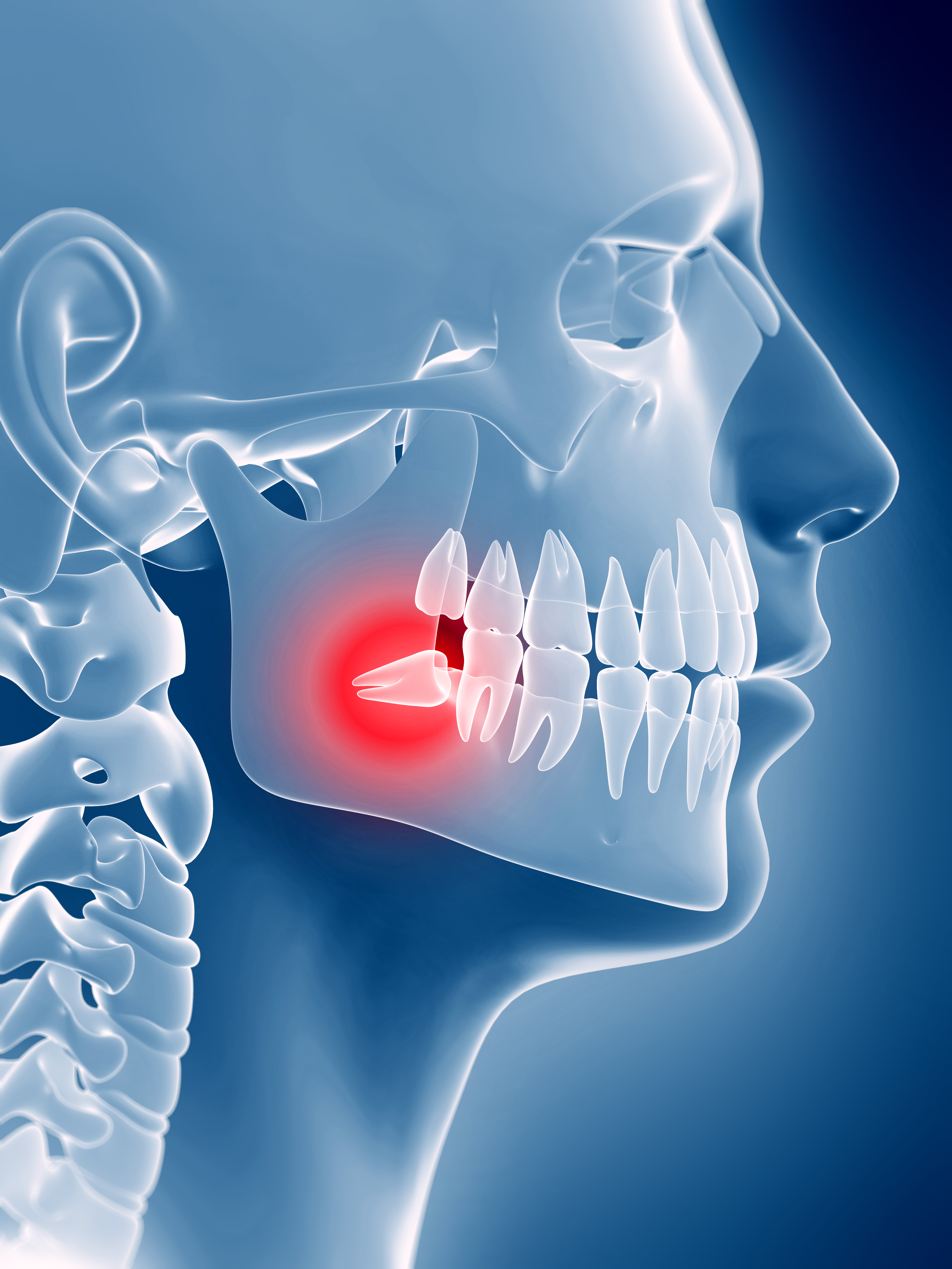
Intra-oral images and X-rays — captured, stored and annotated inside the patient chart. metaDENT Dental Imaging integrates directly with intra-oral sensors, TWAIN-compatible scanners and cameras so clinicians never leave the patient record to work with images.

DICOM native
plus JPG, BMP, TIFF
2 or 4 images
side-by-side comparison
In-chart access
no separate application
3D dental X-ray skull
Dental imaging

Every X-ray and intra-oral photo — linked to the tooth, the chart, the record.

Benefits

What your clinical teams gain on day one.

In-chart image access

Images are opened directly within the dental chart — no switching to a separate imaging workstation or application. The clinician sees the X-ray and the odontogram in the same workflow, at the same time.

Side-by-side comparison

Compare 2 or 4 images simultaneously in a split-screen layout. Monitor periapical healing, track bone levels over successive radiographic examinations, and demonstrate clinical progress to the patient in a single view.

Full image manipulation

Zoom, flip, rotate, and apply brightness, contrast, sharpening and invert filters directly on the stored image. Bring out diagnostic detail without any external image editing software.

Device integration

Direct capture from intra-oral sensors and TWAIN-compatible scanners and cameras with no intermediate steps. Images land in the patient chart the moment acquisition is complete — no import, no manual filing.

Annotate directly

Add freehand drawing, text labels and clinical icon overlays directly on stored images. Highlight pathology, mark planned procedure sites and add notes that remain associated with the image in the permanent record.

Format support

DICOM, JPG, BMP and TIFF — all archiving-ready from day one. Legacy images from prior systems can be imported and linked to the patient dental chart without reformatting or losing metadata.

Feature highlights

Complete dental image management — acquisition to archive.

Chart integration

  • Images linked directly to the affected tooth on the odontogram
  • Accessible from within the dental chart without navigating away
  • Visit-dated image sets for chronological review of any tooth
  • Images visible to authorised clinical staff across the practice network

Image manipulation

  • Zoom in/out with pan and magnifying glass tools
  • Flip horizontal / vertical and free rotation
  • Brightness, contrast, sharpening and invert filters
  • Original image always preserved — filters are non-destructive

Device interfacing

  • Direct acquisition from intra-oral sensors (USB and wireless)
  • TWAIN-compatible scanner and camera interface
  • Automatic patient and tooth metadata on captured images
  • Panoramic and cephalometric import from OPG units

Annotation tools

  • Freehand drawing with configurable line colour and thickness
  • Text label placement at any position on the image
  • Clinical icon overlays (arrows, circles, pathology markers)
  • Annotations stored with the image and printed on referral letters

Comparison viewer

  • 2-up and 4-up split-screen comparison layouts
  • Images from any visit date selectable for each panel
  • Linked zoom — zoom both panels simultaneously for direct comparison
  • Print multi-image comparison views for patient education

Access control & archiving

  • Role-based access control — lock image records from modification
  • Full audit trail of image access, annotation and download
  • DICOM, JPG, BMP and TIFF formats — archiving-compliant from day one
  • Configurable retention policy aligned to local regulatory requirements
Part of metaDENT

Dental Imaging is one integrated module within metaDENT — images, chart, record and billing all connected in one platform.

Dental imaging in metaDENT is not a standalone application bolted alongside the clinical system — it is embedded within the patient dental chart. An X-ray captured from an intra-oral sensor is immediately associated with the patient record, the relevant tooth on the odontogram, and the visit encounter. The dentist never leaves the chart to view, manipulate or annotate an image.

Because images are stored in the same platform as the rest of the patient record, they are available to authorised clinicians anywhere in the network. Referral letters generated from the chart can include annotated images without any additional export step. Images are retained according to the same configurable policy that governs all patient records, ensuring consistent compliance across the practice.